Knowledge Base
Browse articles, videos, and guidelines by specialty.

Wound Healing — Phases
Wound Healing
Key Points 4 phases: Haemostasis → Inflammation → Proliferation → Remodelling Haemostasis: platelet plug, fibrin clot, vasoconstriction. Immediate. Inflammation (days 1–4): neutrophils first (bactericidal), then macrophages (essential — orchestrate healing, debride, release growth factors PDGF, TGF-β) Proliferation (days 4–21): fibroblasts lay collagen III, angiogenesis, wound contraction (myofibroblasts), epithelialisation Remodelling (day 21 → 2 years): collagen III → collagen I, tensile strength increases. Max strength = 80% of original Wound never regains >80% original tensile strength Exam Traps Macrophage depletion → severely impaired healing Collagen type III (healing) vs type I (normal skin, tendon, bone) Epithelialisation occurs from wound edges AND adnexal structures (hair follicles)
Wound Healing — Factors Affecting
Wound Healing
Local Factors Infection (>10⁵ organisms/g tissue = clinical infection) Ischaemia / poor perfusion Foreign body, haematoma, dead space Radiation — damages vasculature, fibroblasts, permanent effect Excessive tension Systemic Factors Diabetes — impaired neutrophil function, microangiopathy, neuropathy Malnutrition — especially zinc, Vit C (collagen synthesis), Vit A (epithelialisation) Steroids — impair inflammation and proliferation (Vit A can partially reverse this) Jaundice, uraemia, malignancy Age — delayed but not fundamentally impaired Exam Traps Vitamin C deficiency → scurvy → impaired hydroxylation of proline/lysine → weak collagen Zinc deficiency → impaired proliferation phase Steroids given AFTER wound is 4 days old have less impact on healing
Skin Grafts — Classification & Physiology
Skin Grafts
Types Split thickness skin graft (STSG): epidermis + part of dermis. Donor site heals spontaneously Full thickness skin graft (FTSG): epidermis + full dermis. Less contraction, better cosmesis, donor must be closed Graft Take — 3 Phases 1. Plasmatic imbibition (0–48h): graft absorbs wound fluid passively — no blood supply yet 2. Inosculation (48–72h): donor/recipient capillaries align 3. Revascularisation (72h+): new vessel ingrowth Why Grafts Fail Haematoma/seroma — most common. Lifts graft off bed Infection Shear — movement disrupts new vessels Poor recipient bed (bare bone/tendon/cartilage without periosteum/perichondrium) Contraction Primary contraction: immediate elastic recoil (more with FTSG — more dermis) Secondary contraction: myofibroblast-driven (more with STSG — less dermis to resist) Exam Traps STSG contracts MORE in the long term (secondary) FTSG recoils MORE immediately (primary) Pie-crusting / meshing increases surface area, improves drainage
Flap Classification
Flap Surgery
By Blood Supply Random pattern: dermal/subdermal plexus. No named vessel. L:W ratio ~1:1 (varies by region) Axial pattern: named artery in long axis. Can be longer Perforator flap: based on musculocutaneous or septocutaneous perforators By Composition Cutaneous, fasciocutaneous, myocutaneous, osseous, osteocutaneous, visceral Mathes & Nahai Muscle Classification Type I: single dominant pedicle (e.g. tensor fascia lata) Type II: dominant + minor pedicles (e.g. gracilis, soleus) Type III: two dominant pedicles (e.g. gluteus maximus, rectus abdominis) Type IV: segmental pedicles (e.g. sartorius) — unreliable for transposition Type V: one dominant + secondary segmental (e.g. latissimus dorsi, pec major) By Movement Advancement, rotation, transposition, interpolation, free Exam Traps Latissimus dorsi = Type V (dominant thoracodorsal + secondary intercostals) Gracilis = Type II — workhorse for functional muscle transfer Rectus abdominis = Type III — DIEP flap based on deep inferior epigastric perforators
Burns — Assessment & Resuscitation
Burns
TBSA Estimation Rule of Nines (adults): head 9%, each arm 9%, each leg 18%, anterior trunk 18%, posterior trunk 18%, perineum 1% Lund & Browder: more accurate, accounts for age-related changes in children Patient's palm (including fingers) = 1% TBSA Burn Depth Superficial epidermal: erythema only, no blisters, painful. Not included in TBSA Superficial partial thickness: blistered, moist, painful, blanches → heals <14 days Deep partial thickness: mottled, reduced sensation, may not blanch → needs surgery Full thickness: leathery, painless, does not blanch → always needs surgery Parkland Formula 4 mL × weight (kg) × %TBSA burned = total Hartmann's in 24h Half in first 8h (from time of BURN, not arrival), half in next 16h Urine output target: 0.5 mL/kg/h adults, 1 mL/kg/h children Referral Criteria (to Burns Unit) >10% TBSA (adult), >5% (child) Full thickness any size Face, hands, feet, genitalia, perineum, major joints Inhalation injury Chemical/electrical burns Circumferential burns Exam Traps Parkland calculates from TIME OF BURN not time seen Superficial epidermal burns NOT included in TBSA calculation Electrical burns: entry/exit wounds underestimate deep tissue damage
Melanoma — Diagnosis & Staging
Skin Cancer
Diagnosis ABCDE: Asymmetry, Border irregularity, Colour variation, Diameter >6mm, Evolution Excision biopsy preferred (2mm margins) — never shave biopsy Breslow thickness = distance from granular layer to deepest tumour cell (mm) AJCC 8th Edition Staging T stage: based on Breslow thickness + ulceration - T1a: <0.8mm, no ulceration - T1b: <0.8mm with ulceration OR 0.8–1.0mm - T2: 1.01–2.0mm - T3: 2.01–4.0mm - T4: >4.0mm N stage: nodal involvement (SLNB) M stage: distant metastasis (LDH important) Subtypes Superficial spreading: most common (70%) Nodular: vertical growth, aggressive, poor prognosis Lentigo maligna: sun-damaged skin, elderly, slow growing Acral lentiginous: palms/soles/subungual, more common in darker skin Desmoplastic: neurotropic, high local recurrence Exam Traps Clark level NO longer used in AJCC 8th edition for staging Ulceration upstages tumour SLNB offered if Breslow >0.8mm or any T1b Desmoplastic melanoma has high local recurrence → wider margins
BCC — Classification & Management
Skin Cancer
Subtypes Nodular: most common, pearly papule with telangiectasia, rolled edge Superficial: flat, erythematous, multifocal — treat with topical or PDT Morphoeic (sclerosing): scar-like, ill-defined margins, highest recurrence — treat with Mohs Infiltrative: aggressive growth pattern Basosquamous: hybrid — behaves more like SCC, can metastasise Management Surgical excision: 3–4mm margins for low-risk, 5–6mm for high-risk Mohs micrographic surgery: best for morphoeic, recurrent, facial, large BCCs Non-surgical: imiquimod, PDT, radiotherapy (elderly/unfit patients) Vismodegib (Hedgehog pathway inhibitor): locally advanced or metastatic BCC High-Risk Features Morphoeic/infiltrative subtype Size >2cm Location: face, ear, scalp Recurrent Perineural/perivascular invasion Immunosuppressed patient Exam Traps BCC metastasises extremely rarely (<0.1%) — basosquamous variant can Gorlin syndrome (PTCH1 mutation): multiple BCCs, jaw cysts, bifid ribs Vismodegib targets Smoothened in Hedgehog pathway
Flexor Tendons — Anatomy & Zones
Flexor Tendons
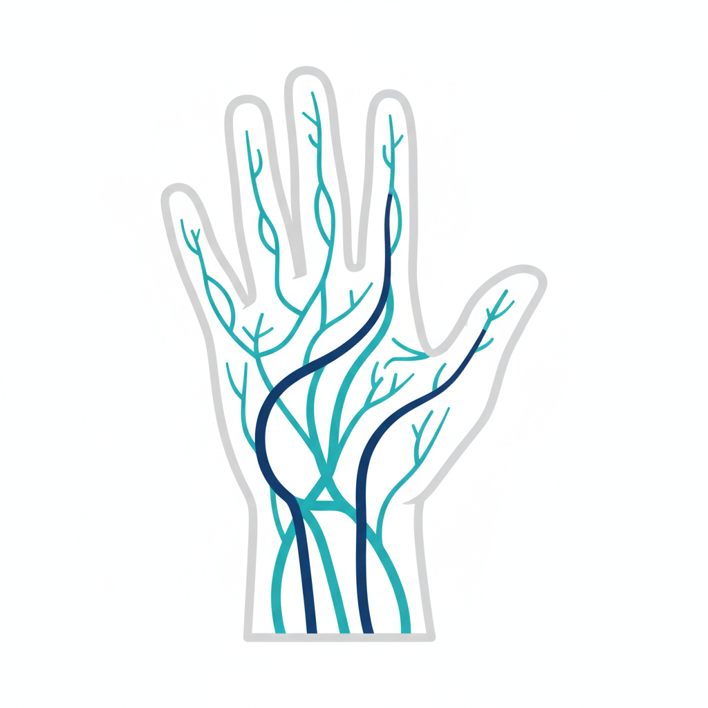
Zone Classification (Verdan) Zone I: distal to FDS insertion (FDP only) Zone II: 'No man's land' — A1 pulley to FDS insertion. Both FDP + FDS in sheath. Highest risk of adhesions Zone III: palm, from carpal tunnel to A1 pulley. Lumbricals originate here Zone IV: carpal tunnel Zone V: forearm, proximal to carpal tunnel Thumb zones: TI (distal to IP joint), TII (A1 to IP), TIII (thenar eminence) Pulley System Annular pulleys A1–A5, Cruciate C1–C3 A2 (proximal phalanx) and A4 (middle phalanx) — most critical. Preserve at all costs A1 pulley release — trigger finger Bowstringing = loss of A2 + A4 Blood Supply to Tendons Vincula longa and brevia — mesotenon extensions carrying blood supply Avascular zones rely on synovial diffusion Exam Traps Zone II injuries have worst prognosis for adhesions FDS inserts as two slips into middle phalanx (Camper's chiasm), FDP passes through Lumbricals originate from FDP in palm — hence paradoxical extension with lumbrical-plus deformity after FDP division
Carpal Tunnel Syndrome
Nerve Compression
Anatomy Median nerve compressed under flexor retinaculum Contents of carpal tunnel: median nerve + 9 tendons (4 FDS, 4 FDP, FPL) Palmar cutaneous branch arises PROXIMAL to tunnel → not affected → preserved sensation over thenar eminence Clinical Features Paraesthesia/numbness: thumb, index, middle, radial half of ring (median distribution) Night symptoms (waking, shaking hand) Thenar wasting (LOAF muscles — except palmaris brevis): Lumbricals 1&2, Opponens pollicis, Abductor pollicis brevis, Flexor pollicis brevis Weakness of thumb opposition and abduction Tests Phalen's: wrist flexion 60 seconds → symptoms Tinel's: percussion over tunnel → tingling Durkan's (carpal compression test): most sensitive/specific NCS/EMG: confirms diagnosis, grades severity Management Non-surgical: night splint, steroid injection (diagnostic + therapeutic) Surgical: carpal tunnel decompression — division of flexor retinaculum - Open or endoscopic (quicker recovery, same outcomes long-term) Exam Traps Thenar sensation PRESERVED (palmar cutaneous branch) APB (abductor pollicis brevis) = best muscle to test for median nerve motor function at wrist Recurrence after surgery: check for incomplete release, secondary causes (synovitis, ganglion)
Nerve Injury — Classification
Nerve Injury
Seddon Classification Neurapraxia: conduction block, no axonal loss. Full recovery. (e.g. Saturday night palsy) Axonotmesis: axonal disruption, endoneurium intact. Wallerian degeneration. Good recovery at 1mm/day Neurotmesis: complete nerve division, no recovery without surgery Sunderland Classification Grade I: neurapraxia Grade II: axonotmesis — endoneurium intact → spontaneous recovery Grade III: endoneurium disrupted, perineurium intact → partial recovery expected Grade IV: only epineurium intact → poor spontaneous recovery, likely needs surgery Grade V: complete transection (neurotmesis) Grade VI (Mackinnon): mixed pattern across fascicles Wallerian Degeneration Distal axon degenerates within 24–72h Schwann cells form Büngner bands — tubes guiding regenerating axons Denervated muscle atrophies — becomes irreversibly fibrosed after ~18–24 months Nerve Regeneration ~1mm/day (3cm/month) distal to injury after 3–4 week latent period Tinel's sign advances distally as regeneration progresses Exam Traps Grade III injury: endoneurium disrupted → axons may regenerate into wrong tubes → incomplete recovery Time is critical — delayed repair has worse outcomes due to target organ changes Tinel's sign advancing = active regeneration; stationary Tinel's = stalled recovery
Dupuytren's Disease
Dupuytren's Disease
Pathology Fibroproliferative disorder of palmar fascia Myofibroblast proliferation → collagen III → collagen I → cord formation Normal fascial structures become pathological cords: pretendinous, spiral, lateral, retrovascular, Grayson's ligament Spiral cord: most complex — displaces NVB towards midline and superficially (risk of injury) Associations (Dupuytren's Diathesis) Northern European descent, male (10:1), age >50 Diabetes, epilepsy (phenobarbitone), alcoholic liver disease, HIV Bilateral, ectopic disease (Garrod's knuckle pads, Peyronie's, Ledderhose) = diathesis = higher recurrence Surgical Indications (Tubiana/Hueston table-top test) MCP ≥30° flexion, OR PIP any flexion contracture (PIP harder to correct), OR Positive table top test (cannot place palm flat on table) Treatment Options Fasciotomy: needle or open — divide cord, no excision. High recurrence Fasciectomy: regional (excise cord + involved fascia) — standard Dermofasciectomy: excise skin + fascia, FTSG — lowest recurrence (used for recurrent disease) Collagenase injection (Xiaflex): clostridial collagenase — less invasive, works best for MCP cords Exam Traps PIP contracture: harder to correct, worse outcomes than MCP Spiral cord → NVB displaced medially and superficially — HIGH injury risk Dermofasciectomy for recurrence or diathesis Garrod's pads + bilateral = diathesis = counsel for recurrence
Anatomy — Fascial Layers of the Hand
Hand Anatomy
Palmar Fascia Central: triangular, continuous with palmaris longus Longitudinal pretendinous bands → digital slips → Grayson's (palmar) and Cleland's (dorsal) ligaments Hypothenar and thenar fascia lateral Natatory ligament: superficial transverse ligament of palm at web spaces Septa Medial and lateral septa divide palm into 3 compartments: thenar, central, hypothenar Central compartment: flexor tendons, lumbricals, neurovascular bundles Dorsal Fascia Dorsal subaponeurotic space: between extensor tendons and interossei Infection can track here from web spaces Fibrous Flexor Sheath Annular (A1–A5) and cruciate (C1–C3) pulleys A2 and A4: biomechanically critical — preserve during surgery Digital synovial sheath: from A1 to DIP level (thumb sheath communicates with radial bursa) Ulnar bursa communicates with little finger sheath Exam Traps Cleland's ligament: DORSAL to NVB (does NOT contract in Dupuytren's) Grayson's ligament: VOLAR to NVB (involved in Dupuytren's spiral cord) Radial + ulnar bursae communicate in 80% → horseshoe abscess if both infected Natatory ligament: web space tightness in Dupuytren's
Anatomy — Intrinsic Muscles of the Hand
Hand Anatomy
Thenar Muscles (Median nerve — recurrent branch) Abductor pollicis brevis (APB): abducts thumb — best test for median nerve Opponens pollicis: rotates MC1 into opposition Flexor pollicis brevis: superficial head (median), deep head (ulnar) Hypothenar Muscles (Ulnar nerve) Abductor digiti minimi, Flexor digiti minimi, Opponens digiti minimi Palmaris brevis: wrinkles hypothenar skin (ulnar nerve, superficial branch) Lumbricals 4 total. Origin: FDP tendons in palm. Insert: radial lateral band Action: MCP flexion + IP extension (intrinsic plus position) Lumbrical 1+2: median nerve. Lumbrical 3+4: ulnar nerve Interossei 4 dorsal (DAB — Dorsal ABduct). 3 palmar (PAD — Palmar ADduct) All ulnar nerve (deep branch) Dorsal interossei: largest — fill dorsal spaces, tested as FDI Action: MCP flexion, IP extension (same as lumbricals) Adductor Pollicis Two heads: oblique (capitate) and transverse (MC3) Deep branch ulnar nerve Action: adducts thumb — Froment's sign when weak Exam Traps Intrinsic plus: MCP flexed, IPs extended — position of lumbricals/interossei action Intrinsic minus (claw): MCP hyperextended, IPs flexed — ulnar nerve palsy FPB has DUAL innervation — may preserve partial thenar bulk in median palsy Lumbrical plus deformity: FDP cut distal to lumbrical origin → active flexion causes paradoxical IP extension
Anatomy — Neurovascular Supply of the Hand
Hand Anatomy
Arterial Supply Radial artery: enters hand via anatomical snuffbox → deep palmar arch (mainly radial) Ulnar artery: superficial palmar arch (mainly ulnar) Superficial arch: distal to deep — gives common digital arteries → proper digital arteries Deep arch: gives princeps pollicis, radialis indicis, palmar metacarpal arteries Allen's test: assesses dominant supply — compress both, release one Venous Drainage Dorsal venous arch → cephalic (radial) and basilic (ulnar) veins Palmar veins less prominent Sensory Distribution Median: radial 3½ digits (palmar), dorsal tips of same digits Ulnar: ulnar 1½ digits (palmar and dorsal) Radial: dorsal radial 3½ digits (proximal — tips are median) Palmar cutaneous branch of median: thenar skin (exits proximal to carpal tunnel) Dorsal branch of ulnar: dorsal ulnar hand (exits 5cm proximal to wrist) Digital Neurovascular Bundles Proper digital arteries and nerves run on either side of each digit Nerve: dorsal to artery at base, volar to artery at fingertip Exam Traps Dorsal branch of ulnar nerve: exits 5cm proximal to wrist — spared in Guyon's canal injuries Radial nerve: ONLY supplies skin — no intrinsic hand muscles Allen's test: positive (abnormal) = dominant artery occluded, hand blanches Princeps pollicis from deep arch — critical for thumb replantation planning

Examinations — FRCS (Plast.) Viva Format
Exam Technique
Format Written: MCQ + EMQ paper (SBA format, single best answer) Clinical/Viva: 4 stations × 25 minutes - Operative surgery station - Clinical cases station - Communication/ethics station - Academic/research station What Examiners Want Safe decision-making — not encyclopaedic knowledge Logical, structured approach to clinical problems Demonstration that you won't harm a patient Evidence-based justification for decisions Clear communication — answer the question asked Common Viva Structures History/assessment → 'What would you do next?' Complications → 'How would you manage this?' Anatomy → Draw it / describe relations Evidence → 'What is the evidence for X?' Approach to Questions Structure your answer: Definition → Classification → Management Offer your view: 'In my practice I would…' If unsure: acknowledge, reason through logically Don't bluff — examiners can tell, and it's a red flag Topics That Always Come Up Flap physiology, wound healing, burns resuscitation Melanoma margins and SLNB Free flap monitoring and salvage Nerve injury classification Skin cancer management pathways Exam Traps 'Tell me about X' = they want you to structure, not free-associate Always mention safety netting / follow-up / MDT Research station: know at least one RCT and one systematic review in your subspecialty Ethics: four pillars — autonomy, beneficence, non-maleficence, justice
Examinations — Hand Diploma Viva Format
Exam Technique
BSSH Hand Diploma Format Written: MCQ paper Oral: 4 × 20 minute viva stations - Trauma (fractures, tendons, nerves) - Elective (Dupuytren's, arthritis, compression neuropathies) - Microsurgery/replantation - Paediatric / congenital hand Core Topics by Station Trauma: Zone II flexor tendon repair, rehabilitation protocols Scaphoid fracture — assessment to management Distal radius fractures — surgical vs conservative Extensor tendon zones and deformities (mallet, boutonnière) Elective: Dupuytren's — indications, techniques, recurrence Carpal tunnel, cubital tunnel — grading and surgical options Rheumatoid hand — deformities and reconstructive options Osteoarthritis thumb CMC — grading, trapeziectomy ± LRTI Microsurgery: Replantation — indications, order of repair Digital nerve repair vs grafting Free functional muscle transfer Paediatric: Syndactyly, polydactyly, radial dysplasia Thumb hypoplasia — Blauth classification and pollicisation Exam Traps Examiners test clinical judgment — 'what would YOU do?' not textbook Know your units' protocols — justify them with evidence Rehabilitation: know at least one protocol per major tendon zone CRPS: know diagnostic criteria (Budapest) and management ladder
MRCS Anatomy — Axilla & Brachial Plexus
MRCS Anatomy
Boundaries of the Axilla Apex: clavicle, 1st rib, scapula Anterior wall: pec major + minor Posterior wall: subscapularis, teres major, latissimus dorsi Medial wall: serratus anterior (ribs 1–4) Lateral wall: intertubercular groove of humerus Floor: axillary fascia + skin Contents Axillary artery (3 parts relative to pec minor) Axillary vein (medial to artery) Brachial plexus cords + branches Axillary lymph nodes (levels I–III relative to pec minor) Long thoracic nerve (C5/6/7 — serratus anterior) Thoracodorsal nerve (C6/7/8 — latissimus dorsi) Axillary Artery Branches (Screw The Lawyers Save A Patient) Part 1 (medial to pec minor): Superior thoracic Part 2 (behind pec minor): Thoracoacromial, Lateral thoracic Part 3 (lateral to pec minor): Subscapular, Anterior circumflex humeral, Posterior circumflex humeral Axillary Lymph Nodes Level I: lateral to pec minor (anterior, posterior, lateral groups) Level II: behind pec minor (central group + interpectoral/Rotter's nodes) Level III: medial to pec minor (apical group) → drains to subclavian trunk Exam Traps Long thoracic nerve: runs on serratus anterior (medial wall) — at risk in axillary dissection → winging Thoracodorsal nerve: posterior wall — sacrificed in radical clearance Level III nodes: apical — clearance causes lymphoedema risk Axillary vein: always MEDIAL to artery
MRCS Anatomy — Shoulder Joint
MRCS Anatomy
Joint Type Ball and socket — synovial. Most mobile, least stable joint in body Glenoid labrum deepens socket by 50% Reinforced by: rotator cuff, glenohumeral ligaments, long head biceps Rotator Cuff (SITS) Supraspinatus: abduction (initiates first 15°), C5/6, suprascapular nerve Infraspinatus: lateral rotation, C5/6, suprascapular nerve Teres minor: lateral rotation, C5/6, axillary nerve Subscapularis: medial rotation, C5/6, upper + lower subscapular nerves Supraspinatus: most commonly torn (critical zone — avascular area 1cm from insertion) Shoulder Abduction 0–15°: supraspinatus initiates 15–90°: deltoid (axillary nerve C5) + supraspinatus 90–150°: scapular rotation (serratus anterior + trapezius) 150–180°: lateral flexion of spine Axillary Nerve From posterior cord (C5/6) Passes through quadrangular space (teres minor, teres major, long head triceps, humerus) Supplies: deltoid + teres minor + skin over deltoid (regimental badge area) At risk: anterior shoulder dislocation, surgical neck of humerus fracture Quadrangular Space vs Triangular Space Quadrangular: axillary nerve + posterior circumflex humeral artery Triangular (medial): circumflex scapular artery only Triangular (lateral/triangular interval): radial nerve + profunda brachii Exam Traps Axillary nerve: regimental badge area numbness after shoulder dislocation Supraspinatus critical zone: 1cm from insertion — watershed area Scapular rotation (not just glenohumeral) allows >90° abduction Long head biceps: intra-articular but extrasynovial
MRCS Anatomy — Inguinal Region
MRCS Anatomy
Inguinal Canal Length: ~4cm, runs superomedially from deep to superficial ring Deep ring: defect in transversalis fascia — lateral to inferior epigastric vessels Superficial ring: defect in external oblique aponeurosis — above pubic tubercle Walls Anterior: external oblique aponeurosis (+ internal oblique laterally) Posterior: transversalis fascia (+ conjoint tendon medially) Roof: arching fibres of internal oblique + transversus abdominis Floor: inguinal ligament + lacunar ligament medially Contents Male: spermatic cord (vas deferens, testicular artery, pampiniform plexus, cremasteric artery, genital branch of genitofemoral nerve, sympathetics, lymphatics) Female: round ligament of uterus Both: ilioinguinal nerve (exits through superficial ring — NOT in cord) Direct vs Indirect Hernia Indirect: through deep ring, lateral to inferior epigastric vessels → congenital (patent processus vaginalis) Direct: through Hesselbach's triangle, medial to inferior epigastric vessels → acquired (muscle weakness) Hesselbach's triangle: inferior epigastric vessels (lateral), inguinal ligament (inferior), rectus sheath (medial) Femoral Canal Below inguinal ligament, medial to femoral vein Contains: lymphatics (Cloquet's node) and fat Femoral hernia: more common in women, high risk of strangulation (rigid neck) Exam Traps Ilioinguinal nerve: exits superficial ring — not inside spermatic cord Direct hernia: medial to inferior epigastric (weakness) — rarely strangulates Indirect hernia: lateral — can descend into scrotum Femoral hernia: below AND lateral to pubic tubercle (inguinal = above and medial) Pantaloon hernia: both direct + indirect components
FRCS Orth — Viva Format & Exam Technique
FRCS Orth Exam
FRCS (Orth) Examination Structure Part 1: MCQ + EMQ (basic sciences) Part 2: Clinical (OSCEs, vivas, short cases) Viva format: 4 viva stations × 20 minutes each Stations: Adult Elective, Trauma, Children's Orthopaedics, Applied Basic Science Marking Each viva: scored 4–8 per examiner (two examiners per station) Pass threshold: 6/8 per station overall Clear fail: 4 | Borderline fail: 5 | Pass: 6 | Good pass: 7 | Outstanding: 8 Viva Approach 1. Listen carefully — state what you see/what you'd do first 2. Structure your answer (history → examination → investigation → management) 3. Prioritise safety: don't skip neurovascular assessment in trauma 4. Use eponyms correctly but explain them 5. If shown an X-ray: describe systematically (AP/lateral, patient details, alignment, bone, cartilage, soft tissue) X-ray Presentation Framework (ABCDS) Alignment Bone density and cortex Cartilage (joint space) Dense shadows (foreign bodies, calcification) Soft tissues Common Viva Traps Being vague about margins/surgical thresholds — quote numbers Forgetting to mention consent and complications Not knowing classification systems (Neer, Garden, AO) Underselling your experience — say 'I would' not 'one would' Key Classifications to Know Cold Fractures: AO/OTA, Garden (NOF), Neer (proximal humerus), Gartland (supracondylar) Soft tissue: Gustilo-Anderson (open fractures), Mason (radial head) Spine: ASIA, Denis (thoracolumbar), AO Spine OA: KL grading (knee), Tonnis (hip)
FRCS Orth — Hip Anatomy & Surgical Approaches
FRCS Orth Anatomy
Hip Joint Ball and socket: synovial, multiaxial Acetabulum: formed by ilium (40%), ischium (40%), pubis (20%) Labrum deepens socket — contains type I and III collagen Zona orbicularis: circular fibres of capsule at femoral neck Blood Supply to Femoral Head Primary: medial femoral circumflex artery (MFCA) — via retinacular vessels Minor: lateral femoral circumflex, obturator (foveal artery — negligible in adults) MFCA at risk in: intracapsular NOF fractures, posterior hip dislocation Capsule & Ligaments Iliofemoral (Y ligament of Bigelow): strongest, anterior — resists extension Pubofemoral: inferior — resists abduction and extension Ischiofemoral: posterior — resists internal rotation Surgical Approaches *Posterior (Moore/Southern):* Most common for THR Patient: lateral decubitus Internervous plane: none (splits gluteus maximus — same nerve supply) Structures at risk: sciatic nerve, short external rotators Short ERs divided: piriformis, obturator internus, gemelli, quadratus femoris *Anterolateral (Watson-Jones):* Internervous plane: TFL (sup gluteal n) / gluteus medius (sup gluteal n) — technically same nerve True internervous: between TFL and gluteus medius At risk: superior gluteal nerve (>3.5cm above GT) *Direct Anterior (DAA/Smith-Petersen):* True internervous: sartorius (femoral n) / TFL (superior gluteal n) Advantage: tissue-sparing, early mobilisation At risk: LFCN (meralgia paraesthetica), LFCA *Hardinge (Direct Lateral / Transgluteal):* Splits gluteus medius and vastus lateralis At risk: superior gluteal nerve if extended >5cm proximal to GT Exam Traps MFCA: always the dominant supply in adults Y-ligament (Bigelow): strongest hip ligament Posterior approach: sciatic nerve most at risk DAA: true internervous plane — least muscle damage
FRCS Orth — Osteoarthritis: Pathophysiology & Non-operative Management
Arthroplasty
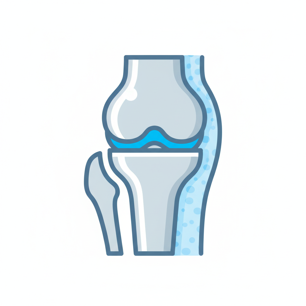
Pathophysiology Progressive cartilage loss + subchondral bone changes + osteophyte formation MMP-mediated degradation of type II collagen + aggrecan IL-1β + TNF-α drive catabolic state Secondary synovitis (not primary — differentiates from RA) KL Grading (Knee) 0: normal | I: possible osteophyte | II: definite osteophyte, possible JSN | III: moderate JSN + sclerosis | IV: severe JSN (<2mm), gross sclerosis Non-operative Management (NICE NG226) 1. Education + self-management 2. Exercise: strengthening + aerobic — first line in ALL patients 3. Weight loss if BMI >25 4. Topical NSAIDs first; oral NSAIDs + PPI; weak opioids 5. Intra-articular steroid: short-term only 6. NOT recommended: glucosamine, arthroscopic washout, PRP Exam Traps Exercise is first line regardless of severity Arthroscopic washout: not recommended for OA IL-1β + TNF-α: key catabolic cytokines KL IV: operative threshold